Using Magnetism to Diagnose Cancers
By: Francis Alcorn
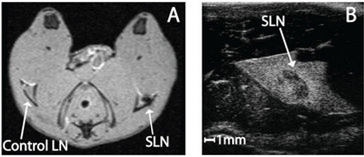
Image taken from Memphis Surgery Associates, PC
Timely treatment is crucial to saving cancer patients’ lives, but to quickly treat patients, doctors must first diagnose the disease and determine how far it has progressed. An important part of the diagnostic process is locating any metastases, pieces which have broken off from the original cancer and travel throughout the body in the lymphatic system. The most likely place to find these metastases is, therefore, in the lymph nodes, especially sentinel lymph nodes, the first lymph nodes which drain a cancer. However, locating where these lymph nodes are can be difficult, slowing diagnosis.
Researchers have been investigating a new method of locating these lymph nodes through the use of superparamagnetic iron oxide nanoparticles. These are tiny bits of iron which interact with magnetic fields, similar to how iron shavings follow a magnet.
To attempt to locate these sentinel lymph nodes, the researchers began by injecting one leg of each of eight rats with nanoparticle mixtures of differing sizes and concentrations, leaving the other leg as the control. Then, the nanoparticles were transported via the lymphatic system to the lymph nodes. Thus, to locate the lymph nodes the researchers looked for areas of high nanoparticle concentration. They did this using two methods; MRI and magneto motive ultrasound imaging, or MMUS. The nanoparticles produced voids in the MRI images, indicating the location of the lymph nodes, but with limited clarity.
On the other hand, the results from the use of MMUS imaging were much more clear and defined. This method involved using magnetic fields to move the nanoparticles. This motion was detected and displayed in the ultrasound image. The resulting picture clearly displayed the lymph node.
 Images taken of the rat lymph nodes in the study. On the left (Image A), is an MRI image of the lymph node, as indicated by the small void in the image. On the right (Image B) is a MMUS image of the lymph node, the dark, bean shaped area in the center.
The researchers also found that the use of smaller nanoparticles at higher concentrations resulted in a better image. They attribute this to be due to smaller particles traveling more easily through the lymphatic systems, such as how a marble will travel more easily through a PVC pipe than a golf ball. For this reason smaller nanoparticles accumulated in the lymph nodes more easily.
Additionally, a higher concentration of nanoparticles injected into the rats meant more particles in total, thus more could accumulate in the lymph nodes. Under the correct experimental conditions, the researchers had very clear ultrasound pictures of the rats’ lymph nodes
While this research does not directly improve cancer treatment as it stands today, it does have significant the potential to accelerate diagnosis, which indirectly improves cancer treatment and increases survivorship.
Works Cited
Evertsson, Maria, Pontus Kjellman, Magnus Cinthio, Sarah Fredriksson, Rene In ‘t Zandt, Hans Persson, and Tomas Jansson. “Multimodal Detection of Iron Oxide Nanoparticles in Rat Lymph Nodes Using Magnetomotive Ultrasound Imaging and Magnetic Resonance Imaging.” IEEE Transactions on Ultrasonics, Ferroelectrics, and Frequency Control 61.8 (2014): 1276-283. IEEE Xplore. Web. 23 Nov. 2014. doi:10.1109/TUFFC.2014.3034
Mahmoudi, Morteza, Shilpa Sant, Ben Wang, Sophie Laurent, and Tapas Sen. “Superparamagnetic Iron Oxide Nanoparticles (SPIONs): Development, Surface Modification and Applications in Chemotherapy.” Advanced Drug Delivery Reviews 63.1-2 (2011): 24-46. ScienceDirect. Web. 22 Nov. 2014.
Mehrmohammadi, M., J. Oh, S. Mallidi, and S. Y. Emelianov. “Pulsed Magneto-motive Ultrasound Imaging Using Ultrasmall Magnetic Nanoprobes.” PMC 10.2 (2011): 102-10. PubMed. Web. 22 Nov. 2014.
“Memphis Surgery Associates.” Memphis Surgery Associates. Memphis Surgery Associates, 2013. Web. 01 Dec. 2014.